Respiratory System of Frog
Respiratory System of Frog
Respiration is a process in which food are oxidized with oxygen in order to release energy. The released energy is utilized to perform various life activities. The metabolic waste like CO2 is eliminated from the body.
C6H12O6 6CO2 + 6H2O +energy
Due to amphibious mode of life, frog shows different modes of respiration. The exchange of gaseous takes place in following four ways:
- Gill respiration
- Cutaneous respiration
- Bucco-pharyngeal respiration
- Pulmonary respiration
-
Gill respiration:
In tadpole condition, such type of respiration takes place through 4-5 pairs of gills. The gills are distributed with blood vessels and absorb oxygen through diffusion. The oxygen combines with hemoglobin of blood and forms oxyhaemoglobin which goes to tissue.
- Cutaneous respiration:
The respiration which takes place through skin is called cutaneous respiration.
It takes place in water and during aestivation and hibernation when metabolism is low and demand for oxygen is small. Skin absorbs oxygen dissolved in water through blood capillaries where oxygen combines with hemoglobin. It is carried into different parts of the body by blood and release energy.
The CO2 produced as a waste product is mixed with hemoglobin and forms carboxy-hemoglobin which goes to veins of skin. Later it is passed outside.
When the skin is completely dries, the cutaneous respiration is not possible. Consequently, the frog dies due to asphyxia.Skin of frog is respiratory in nature because
It is profusely supplied with blood capillaries.
Skin is thin and devoid of any structure that prevent diffusion of dissolved oxygen from water in the blood.
Secretion of mucus by mucous gland always keeps its surface moist.
-
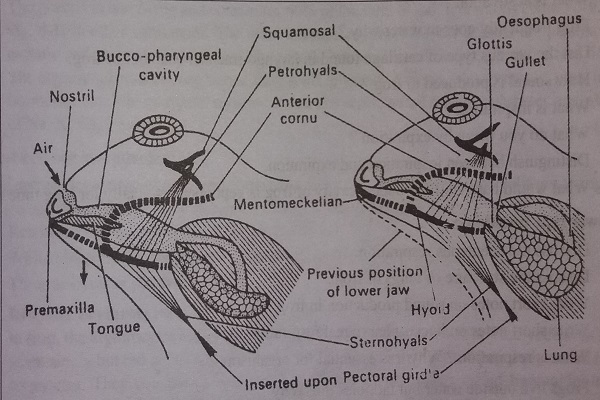
Bucco-pharyngeal respiration
Simply known as mouth respiration.
The respiration which takes place through buccopharyngeal cavity, it is called buccopharyngeal respiration.
The buccal cavity consists of moist mucous membrane and richly supplied with blood capillaries.It absorbs oxygen through diffusion or simply by contraction or expansion of sternohyals and petrohyals muscles. Oxygen dissolves in moist mucous of the cavity and diffuses into the blood capillaries. Similarly, CO2 diffuses out into the cavity and passes out through nares during expiration.
[The lungs donot take part in this respiration.
Sternohyal muscle lies below the mouth cavity. It connects hyoid and sternum when it contracts, the mouth cavity is lowered downwards. The petrohyal muscle lie outside the mouth cavity and the upper and lower ends connect squamosal bone and hyoid respectively. When it contracts, the mouth cavity is pushed upwards.]
Mechanism of bucco-pharyngeal respiration
- Bucco-pharynx or buccal cavity
- Contraction of sternohyal muscles
- Lowering of floor of the cavity
- Area of buccal cavity increases
- Air comes inside the buccal cavity through external nares
- Gas exchange occurs
- Petrohyal muscles contracts
- Raising of floor of buccal cavity
- Area of buccal cavity decreases
- Pressure of air contained increases
- Air goes out through external nares.
-
Pulmonary respiration
It is also known as lung respiration.
Frog respires through lungs when it lives on land.
When frog is more active during locomotion, swimming in water, during leaping and jumping, the demand of oxygen increased.
For pulmonary respiration, the routes of air passages are as follows:
External nares, olfactory chamber, internal nares, buccopharyngeal cavity, glottis, laryngo-tracheal chamber, bronchi and lungs.
At this moment, the cutaneous and buccopharyngeal respiration are not able to supply the desired amount of oxygen and then the frog respire by lungs to fulfill their demand.
Mechanism of pulmonary respiration/ Breathing Mechanism
Breathing is mechanical process. It involves taking air into respiratory organs (inspiration) such as lungs and removing air out (expiration) by changing the volume of bucco-pharyngeal cavity, which acts as a force pump.
Breathing occurs in two steps:
- Inspiration
- Expiration

-
Inspiration
It is the inhalation or intake of fresh air from atmosphere into the lungs for gaseous exchange.
The passage of air from outside into the bucco-pharyngeal cavity is called aspiration.
The process by which lungs are filled with fresh air is called pulmonary inspiration.
Steps occur during inspiration are
- Contraction of sternohyal muscles
- Increase volume of buccopharyngeal cavity
- Decrease pressure of air inside buccal cavity
- Air rushes into the cavity through external nares
- Contraction of submental muscles pushes the mento-mechkelian bones of lower jaw forward and then pushes the premaxillae upward. This closes the external nares.
- Contraction of petrohyal muscles
- Decrease volume of bucco-pharyngeal cavity
- Area decreases and pressure of air increases. This closes the gullet since there is no other outlet for air to escape
- Glottis open
- Air rushes into the lungs and then into alveoli (gaseous exchange occur between blood and alveoli by diffusion)
-
Expiration
The process of removing of air from the lungs to outside is called pulmonary expiration.
It is the exhalation or giving out of carbon dioxide from lungs back to the bucco-pharyngeal cavity through glottis and to outside.
Steps that occur during expiration are given below
- Contraction of lungs and abdominal muscles
- Decrease in volume of lungs
- Pressure increases in lungs
- Contraction of sternohyals muscles
- Lower the floor to increase the volume of cavity
- Decrease pressure of air inside buccal cavity
- Air rushes from lungs into the cavity through glottis
- Relaxation of submental muscles
- Premaxillae come back to its original position
- External nares open
- Contraction of petrohyal muscles
- Decrease volume of bucco-pharyngeal cavity
- Area decreases and pressure of air increases inside the cavity
- Glottis close
- Air rushes outside through external nares
Lungs
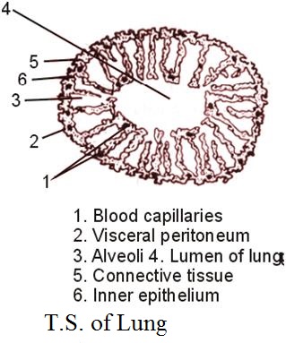
Lungs are a pair of thin walled, oval, hollow, soft and spongy elastic sacs. They are situated in the anterior part of body cavity on each side of the heart. They are pink in color. The wall of lung is made up of three layers. The lung is protected by outermost layer called peritoneum. Below this, there is a connective tissue consisting of blood vessels and muscles fibers. The innermost layer is made up of very thin and flattened ciliated epithelial cells. The inner surface is divided into series of small chambers by irregular septa called alveoli or air sacs. The alveoli greatly increases the surface area to air for gaseous exchange. Lungs are highly vascular and lined with mucus secreting goblet cells. The mucous keep the inner surface of lungs moist for efficient absorption of oxygen.
Respiratory System of Frog