External Morphology of Earthworm
External Morphology of Earthworm
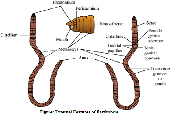
External structures which are visible from outside is called external features or external morphology. The important external features are as follows:

- Shape: Earthworm is elongated, long, narrow, cylindrical or vermiform shaped. The anterior end is tapering while the posterior end is more or less blunt.
- Body surface: Body consists of dorsal and ventral surfaces.
- Size: A mature worm measures about 150 mm in length and 3-5 mm in width.
- Coloration:
i. Color of the body: Dark-brown in color due to the presence of porphyrin pigment which is found in dorsal surface of the body. Dorsal surface is relatively darker than ventral surface.
Porphyrin protects the body from injurious effect of UV-rays.
ii. Respiratory pigment: Hemoglobin which is found in blood plasma that gives bright color of the blood. Hemoglobin is produced by blood gland cells that are present in 4th, 5th and 6th segments.
5. Segmentation: Segmentation of the body is true i.e. external segmentation corresponds to internal segmentation. Hence, the segmentation of earthworm is called metameric segmentation. Body consists of 100-120 segments.
Note: The segments are divided externally by grooves and internally by circular septa.
- Symmetry: Bilaterally symmetrical
NOTE: A type of arrangement of the parts and organs of an animal in which the body can be divided exactly and equally into two halves that are mirror images of each other along one median plane only. - Eyes: No eyes instead of eyes phasomes act as photoreceptor.
- Prostomium and Peristomium: Earthworm lack distinct head. The first segment at the anterior end of the body is called peristomium or buccal segment which bears crescentic mouth.
Peristomium bears a fleshly lobe-like structure is called prostomium which hangs over the mouth ventrally. Prostomium is an aperture which is located in the tip of the first segment. It is called boring part so it bores in soil. - Clitellum:
In mature earthworm, 14th, 15th and 16th segments are enclosed by thick-collar or girdle-like glandular tissue called clitellum.
Cocoon formation takes place in clitellum.
Absent in immature worm hence it indicates the maturity of the worm.
Gland of clitellum secrete mucus, albumen.
It divides the body into 3 region such as:
i. Pre-clitellar region: Starts from 1st to 13th segments
ii. Clitellar region: 14th, 15th and 16th
iii. Post-clitellar region: from 17th up to posterior region.
Clitellum is called Forest of Nephridia.
- Setae: are locomotory organ of earthworm.
Each segment except first, last and clitellum bears setae.
The number of setae are 80-120 per segments.
Each setae is minute, elongated and S-shaped and faint yellow in color.
Each setae consists of 3 parts: Upper parts is neck, Middle swollen part is nodulus and inner part-is root or body, which is attached with setal sac along with muscles.
The clitellar segment possess setae when the worms are immature but setae are shed off before the clitellum is formed at maturity.
The setae are half embedded in the body wall and half projected backwards upon the surface.
NOTE: Setae is formed of horny nitrogenous organic substance known as chitin.
Setae are arranged in annular row in the mid-ventral surface of each segment. This type of arrangement is known as perichaetine arrangement.
- Body wall
Body wall of earthworm is very thin, soft, shiny, elastic and highly vascular. It consists of:
i. Cuticle
It is thin, elastic and non-cellular layer.
It is a layer which is secreted by underlying epidermis.
ii. Epidermis
It is single-layered but multicellular. It lies just below the cuticle. It consists tall and cylindrical epidermal cells which is of following types:
- Supporting cells- These are of columnar type and are major part of epidermis.
- Gland cells- These are thicker consisting of numerous mucous cell and few albumen cells with secretory granules.
The mucous cells are clubbed shaped and secrete mucus while the albumen cells are cylindrical and produce albumen.
- Basal cells- These are small conical cells, lying between supporting cells and gland cells. The cells are later on differentiated into supporting cells and gland cells hence it is also known as replacing cells.
- Sensory receptor cells- These are narrow, columnar cells occurring in groups. These contain hair like processes and sensory in function.
iii. Muscular layer
Muscle layer lies below epidermis.
It consists of an outer circular muscle fibers and an inner longitudinal muscle fibers.
Outer circular muscles consist of numerous scattered granules of porphyrin pigment.
Two additional muscles are also found at the base of each setal sac. They are- A pair of protractor muscles and a single retractor muscle.
iv. Coelomic epithelium
It is the outer envelope of coelomic cavity and hence called coelomic epithelium or parietal peritoneum consisting of flat cells. These flat cells are recognized by nuclei.
Function of Body wall
- It provides definite shape to the body (due to its elasticity).
- Moist body wall helps in respiration.
- Protects the internal delicate organ from injury.
- Secrete mucus which keeps body surface slimy and kills harmful bacteria.
- Alternative contraction and relaxation of circular and inner longitudinal muscle helps in movement.
- Albumen secreted by clitellar gland helps in nutrition of embryo developing inside cocoon.
- Sensory epidermal cells serve for reception of external stimuli.
I need a application to study about earthwarm so if you know about this application plzz suggest me…
thanxxx